

Skeletal model with moving spine, spinal nerves ligaments, etc.
Skeleton model with movable spine, spinal nerves, ligaments, etc.
 Part-pay from 140.62 €/mo
- In stock – delivery 4-8 days
- Cheapest shipping from 26.78 €
- Pay with Swish, card or invoice
- Personal customer service by phone and e-mail
- Swedish online store since 2008
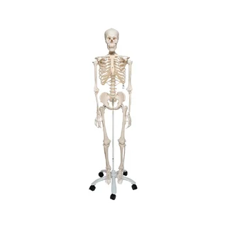
This skeleton model shows the entire skeleton from head to toe in life-size. It also includes the following extra details:
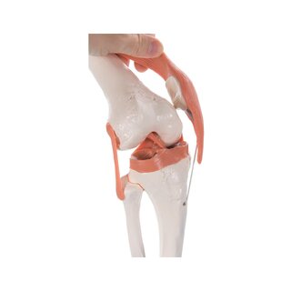
Visible ligaments at the large joints on the right side of the skeleton (e.g. the shoulder joint)
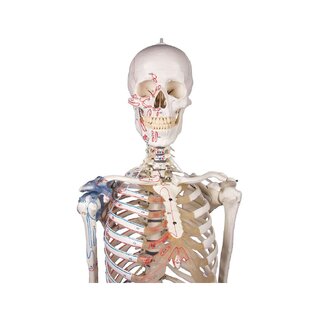
Illustrations of the origin and attachment of skeletal muscles in red and blue on the left side of the skeleton.
Names of muscles and bones
Vertebral arteries
Spinal nerves
A herniated lumbar disc
A highly mobile spine
Mounted on the stand, the plastic skeleton is approx. 177 cm tall, and the level of detail on the legs is high. Most of the joints are held together by metal buckles/metal, which limits the mobility of the joints. The large joints on the right side of the skeleton are held together by brown rubber ligaments and the mobility of these joints is very limited. The spine, however, is very flexible and the mobility is described below.
The skeleton rests on a sturdy stand that has a 5-branched foot with wheels, which makes it easy to move around. A transparent protective bag is included.
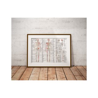
As mentioned above, the origin and attachment of the skeletal muscles are marked on the left side of the skeleton. The bones are also numbered. An overview with the names of bones and muscles is not included in the package when purchased, but can be downloaded here on our website - see under the product images on the left.
Anatomically, the skeleton shows all the bones of the body (including the hyoid bone and sesamoid bone). Cartilage in the upper and lower jaw is also seen. On the left side of the skeleton, the most important ligaments in the shoulder, elbow, hip, and knee joints are seen. The skull can be separated from the rest of the skeleton and divided into three parts (the top of the skull, the lower jawbone and the rest of the skull).
In addition, the vertebral arteries, spinal nerves and a herniated disc in the disc between L3 and L4 (third and fourth lumbar vertebrae) are visible.
The level of detail on the bones is high. Large and small anatomical "landmarks" are shown - for example, the tuberculum majus and minus on the humerus and the linea aspera on the back of the femur (thigh bone). The epiphyseal lines on the upper and lower extremities (arms and legs) are also shown.
In terms of movement, the model is extra flexible in the back, but not in other joints. The joints on the left side of the skeleton are connected with metal buckles/metal that limit the range of movement. For example, flexion and extension (bend and stretch) but not rotation can be seen in the knee joint. The ligaments on the right side of the skeleton severely limit the mobility of these joints.
When the spine is bent, it remains bent until it is straightened again. The spine can perform flexion-extension, lateral flexion and rotation in both the lumbar and thoracic spine. All head movements can be demonstrated in the upper cervical joint, but not in the lower cervical joint (between C1 and C2).
Clinically, this model is developed to demonstrate diseases such as scoliosis, herniated discs and the impact of the spinal nerve roots.The skeleton is also perfect for understanding other diseases where the skeleton plays a role.
| Item number | 1020176 |