

Male pelvis median section
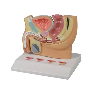
This median section of the male pelvis is with genitals and a removable half bladder and rectum
 Part-pay from 34.76 €/mo
- In stock – delivery 4-8 days
- Cheapest shipping from 11.06 €
- Pay with Swish, card or invoice
- Personal customer service by phone and e-mail
- Swedish online store since 2008
Male pelvis median section
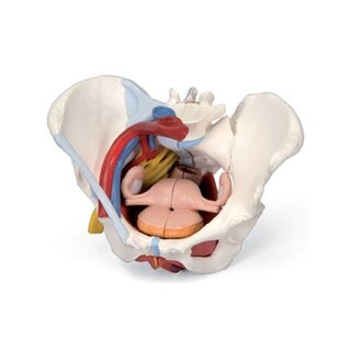
This median section of the male pelvis is with genitals and a removable half bladder and rectum. The other halves are shown in their normal position in the pelvis. The model is delivered on a base with the option of being mounted on the wall.
Anatomically, the model shows many different tissues and organs, some of which can be seen in 3 dimensions, as the model can be separated.
The pelvis (pelvic ring) consisting of bones and the pelvic floor can only be felt on the model. For example, a cross-section of the symphysis (front joint) of the pelvic ring is visible. On the other hand, part of the colon, rectum, a ureter, the great artery and vein in the pelvis and parts of their branches are visible. Ligaments are largely not visible, while nerves are not visible at all (the spinal cord is visible, however).
Some tissues/organs are also visible in the horizontal plane (also called the transverse plane) when looking at the model "from above". Here you can see, among other things, the back and abdominal muscles and the renal tissue in the right kidney. NOTE: All of this cannot be seen in the images on the left but in the PDF file.
The internal genital organs include the testicles and epididymis, which are seen in relation to the scrotum and the spermatic cord (funicle). The contents of the spermatic cord are clearly visible, while this is not the case for its posterior part/blade. In addition, the internal structures of a testicle are visible - but not in fine detail.
The seminal vesicles include the ductus deferens (seminal vesicle), the ductus ejaculatorius and the urethra masculinum, which are all visible (the ductus ejaculatorius can only be felt).
In relation to the accessory gonads, the prostate and vesiculae seminales are seen, but not the glans. bulbourethrales and gll. urethra. The exit passages and their connection can only be felt.
The external genitalia include the penis and scrotum (scrotum), which are also seen. The internal structure of the penis is also seen in small detail.
Many of the pelvic organs function as a channel or reservoir (e.g. the rectum and bladder), and therefore they are seen in a pedagogical way with an air-filled cavity (cavity).
Minimize
In terms of movement, the pelvis is not flexible. The few joints that are visible are not movable.
Clinically, the model can be used to understand
| Item number | H211 |