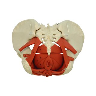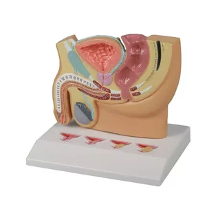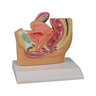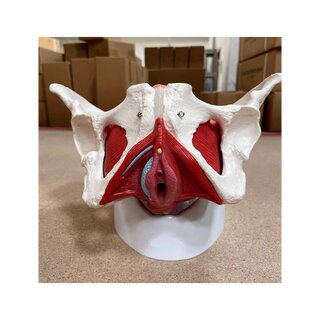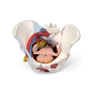


Pelvis model showing bones and joints in the female pelvis
This pelvic model shows the female pelvis and the 2 lower lumbar vertebrae. The model is cast in the natural size of the pelvis, and the level of detail on the legs is good. The joints are assembled with metal pins and nuts.
 Part-pay from 19.99 €/mo
- In stock – delivery 4-8 days
- Cheapest shipping from 17.60 €
- Pay with Swish, card or invoice
- Personal customer service by phone and e-mail
- Swedish online store since 2008
This pelvic model shows the female pelvis and the 2 lower lumbar vertebrae.
The model is cast in the natural size of the pelvis, and the level of detail on the legs is good. The joints are assembled with metal pins and nuts. The front joint (symphysis) is also glued and consists of soft silicone. Via the joints, the pelvis can be separated if desired. Below you can read more about the pelvic dimensions in relation to the pelvis as a mode of delivery.
We sell fetal dolls, which are intended for birth preparation. Our fetal doll with placenta (placenta) and umbilical cord fits exactly to this pelvic model, so that the pelvis as a birth canal can be demonstrated with the doll.
Anatomically speaking
Anatomically, the model shows the female pelvis. Just like in men, the pelvis consists of the 3 building blocks, which are the 2 hip bones and the sacrum. The model also shows the 2 lower lumbar vertebrae. Overall, the model shows the following bones:
Os coxae (the 2 hip bones) of which both are composed
- Os ilium (hip bone/hip bone)
- Os ischii (sit bone)
- Os pubis (pubic bone)
Us sacrum (sacrum) incl. os coccygis (coccyx)
4th and 5th lumbar vertebrae (lumbar vertebra)
The most important details are visible in the bone tissue, such as large tubercles. Smaller details such as the linea glutea are omitted.
The pelvis as a birth canal
Normally, the following 3 connection lines are indicated:
- Conjugata vera (normally measures 11 cm in the female pelvis)
- Diameter obliqua (usually measures 12.5 cm in the female pelvis)
- Diameter transversa (usually measures 13 cm in the female pelvis)
On this pelvic model, the measurements above are: 10.5 cm, 13 cm and 14.5 cm respectively.
You can also use a measurement for the anteroposterior exit from the pelvis, which is the distance from the symphysis to the tip of the coccyx. On this model, the distance is about 11 cm (but it depends on the position of the coccyx, since it is movable).
Movement-wise
In terms of movement, the model's joints should be mentioned. Both the SI joints (articulatio sacroiliaca) and the symphysis (symphysis pubica) are held together by screws and nuts. They are easy to loosen so that the joints can also be "loosened" and moved. The symphysis is also glued. With a firm pull, it can still be loosened (when the nut on the screw is also loosened).
The joint between the sacrum and the coccyx (articulatio sacrococcygea) is extremely flexible, because the coccyx is held firmly on the sacrum with the help of an elastic band (which is not particularly visible). Furthermore, the coccyx consists of 2 parts, which are also movable in relation to each other, because they are also held together with the help of the elastic.
Joints in the 2 lower lumbar vertebrae (facet joints and intervertebral discs) are not movable at all.
The femoral stump variant includes the upper part of the femur bones, mounted on elastic, which allows free movement. The femoral stumps cannot be removed.
Clinically
Clinically, the model can be used in connection with birth preparations and the like. The pelvic model can also be used to understand pelvic fractures with malposition and specific diseases such as axial spondyloarthritis, which among other things involves The SI joint.
< p>
| Item number | A219 |