Classic brain real cast
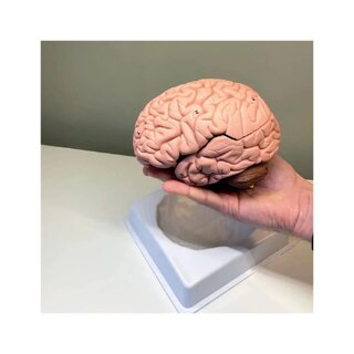
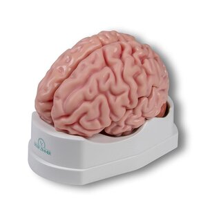
Classic brain, real cast. If you are looking for a brain model that can simply be split down the middle and does not look overly lifelike, this model is ideal.
 Part-pay from 35.75 €/mo
- Cheapest shipping from 11.09 €
- Pay with Swish, card or invoice
- Personal customer service by phone and e-mail
- Swedish online store since 2008
The brain model is cast in hollow, sturdy plastic, which makes the material pliable: it can be squeezed together quite a lot and flexes a little. This makes the model pleasant to handle and work with when it is taken apart and studied.
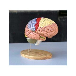
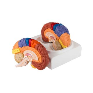
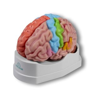
Different areas of the brain are shown in different shades. Below you can read more about anatomical details such as the limbic system. The model can be split down the middle, and the 2 halves are held together by small metal pins. The size of the model corresponds to the brain of an adult. The dimensions are 15 x 14 x 17.5 cm. It weighs approximately 800 grams and comes on a white removable stand.
Important anatomical structures are numbered on the model.
NOTE: The numbering and naming are for guidance only. Please use them critically, as the marker for one anatomical structure may, for example, lie on the border of another structure.
Anatomy
Anatomically, the model shows the human brain, which can generally be divided into the cerebrum, the cerebellum and the brainstem (truncus encephali).
These 3 structures are clearly separated by different colour tones, but the difference between grey and white matter cannot be seen on this model (although it can be seen in the cerebellum).
In the cerebrum (telencephalon and diencephalon), the main features seen are the cerebral lobes, as well as the thalamus and hypothalamus (and the pituitary gland).
In the cerebellum, the vermis cerebelli and the cerebellar hemispheres (hemispherium cerebelli) can be seen.
In the brainstem you can see its 3 parts (midbrain, pons and medulla oblongata) as well as the apparent origins of the cranial nerves.
Other structures can also be seen, such as the fornix, part of the ventricular system and the first 2 cranial nerves (the olfactory and optic nerves), which do not originate from the brainstem.
The limbic system
Many of our customers ask about the limbic system when buying brain models, hence this description.
The limbic system comprises various anatomical structures in the central nervous system (CNS) and is primarily responsible for emotional functions such as anxiety, aggression, mood, memory and social adaptability. Clinically, it is therefore often associated with psychiatric disorders.
The limbic system includes, among others, the amygdala, hippocampus, parahippocampal gyrus, hypothalamus, fornix, mammillary bodies, the prefrontal cortex and the monoaminergic systems in the brainstem. The list is somewhat longer, especially since many fibre connections link the limbic structures. Many customers ask specifically about the amygdala and hippocampus (which is why they are mentioned first in this section).
NOTE: Neither the amygdala nor the hippocampus can be seen on this brain model, but some of the other limbic structures can (e.g. the fornix).
The amygdala is involved in anxiety and the emotional colouring of sensory impressions. It lies as an almond-shaped nucleus IN FRONT OF the hippocampus in the anterior pole of the temporal lobe (the amygdala and hippocampus are therefore separate).
The hippocampus is involved in memory. It lies as an irregular, curved structure in the medial part of the temporal lobe.
For either of these 2 structures to be clearly visible on a brain model, the brain tissue must be shown in a so-called frontal/coronal section through the temporal lobe. The frontal section roughly corresponds to the cutting direction "from ear to ear".
Since the amygdala lies IN FRONT OF the hippocampus (roughly speaking, further "towards the forehead"), both structures can only be seen on a brain model if the model includes at least 2 frontal sections through the temporal lobe, or if the brain model is partly transparent.
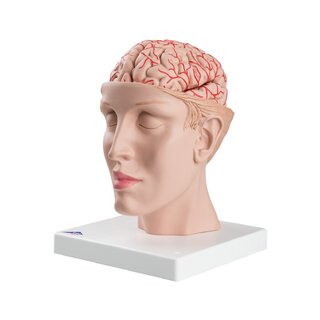
We have not yet seen a brain model that shows 2 sections through the temporal lobe so that both the amygdala and hippocampus are visible. However, our range includes a partly transparent brain model that shows both structures.
All brain models in our range can be separated into different parts. All models (both with and without educational colours) that can be divided into 4 or more parts show the hippocampus. On almost all of these models, the hippocampus is also numbered and named on an overview that can be downloaded from the product descriptions of the brain models.
Clinical use
Clinically, the model can be used for a general understanding of lesions and disorders in the areas of the brain shown on the model.
| Item number | 1000222 |