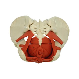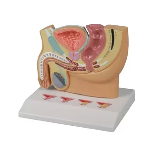


Male brook model in 7 parts
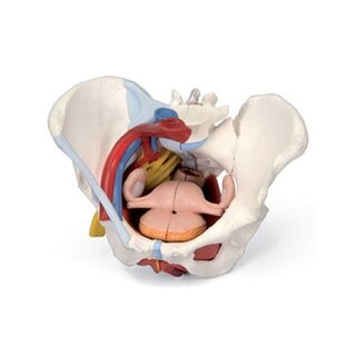
This 7-part model of the male pelvis shows in detail how all the bones, ligaments, blood vessels, nerves, as well as the pelvic floor muscles and the external genitalia are positioned in relation to each other. Right side of the M. sphincter ani externus…
 Part-pay from 104.16 €/mo
- In stock – delivery 4-8 days
- Cheapest shipping from 11.16 €
- Pay with Swish, card or invoice
- Personal customer service by phone and e-mail
- Swedish online store since 2008
The right half of the pelvis shows parts of the V. Iliaca communis, Interna and Externa and shows their position in relation to each other. The sacral plexus, N. ischiadicus, N. pudendus, N. dorsalis penis, NN. scrotalis anteriores, Nn. perinealis and Nn. Annales Inferiores and the ductus deferens are also shown. In addition, the model shows the following bones and ligaments: Both hip bones, symphysis, sacrum and coccyx and L5 with herniated disc. A medial incision is made through L5, sacrum and coccyx so that the entire pelvis can be divided in two. This allows part of the cauda equina to be visible in the spinal canal. The western half of L5 can be removed.
The following ligaments are visible: Lig. inguinale, Lig. sacrotuberale, Lig. sacrospinale, Ligg. sacroiliaca anteriora, Lig. iliolumbale, Lig. longitudinal anterior, Lig. Supraspinale, Lig. sacroiliac interosseum, Lig. sacroiliac posterior, Lig. lateral sacrococcygeum, Lig. sacrococcygeum posterius superficiale et profundum, Membrana obturatoria and Lig. lacunae
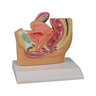
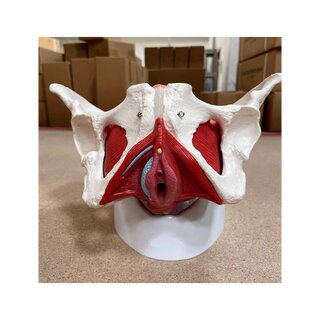
The model is often used together with the female version of the same model with item number: 1000288
| Item number | 1013282 |