Elbow Models
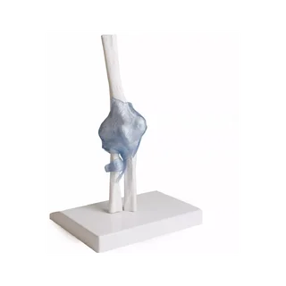
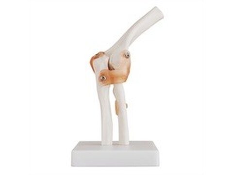
Our range of anatomical elbow models includes both elbow models with and without soft tissues such as ligaments, muscles and nerves.
An elbow model shows the elbow (articulatio cubiti) and thus also the upper arm bone (humerus) as well as the forearm bones called the ulna (elbow) and radius (radius). If the elbow model also shows ligaments, ligaments are typically seen. annular radii, lig. collateral ulnar and / or. collateral radial.One of the elbow models shows the nerves called n. Medianus, n. Ulnaris and n. Radialis, as well as muscles such as with biceps brachii, with brachialis, with flexor carpi radialis and m. Pronator teres. Our most complete elbow model shows both visible muscles and ligaments.
It shows all the muscles belonging to the upper and lower arms. Some of the muscles are directly represented on the model - the remaining muscles are illustrated by the red and blue color of the bones, indicating their origin and attachment.
Clinically, elbow models can be used to understand, for example, tennis and golf elbow, other muscle diseases, fracture (fracture), joint displacement (weakening), sprain and osteoarthritis (osteoarthritis).