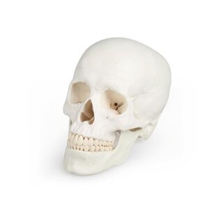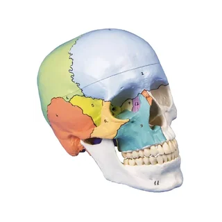


Skull model with many sections and 13 removable parts
A life-size, European original, plastic-cast cranial model with 13 removable parts held together by magnets and pins, making it incredibly easy to disassemble and reassemble the many parts.
 Part-pay from 84.52 €/mo
- In stock – delivery 4-8 days
- Cheapest shipping from 11.18 €
- Pay with Swish, card or invoice
- Personal customer service by phone and e-mail
- Swedish online store since 2008
A life-size, European original, plastic-cast cranial model with 13 removable parts held together by magnets and pins, making it incredibly easy to disassemble and reassemble the many parts.
The skull is particularly interesting for dentists and ENT doctors, who want a model that educationally clears the skull's many complex internal structures and cavities.
Anatomically, the skull shows the following
The cranial tip is removable, but leaves the temporal bones and their sutures intact. Bone impressions of the sagittal, transverse and sigmoid sinuses as well as the meninges are visualized with color. The skull base is cut sagittally in such a way that the cut goes through a sieve plate on one side and another is cut with the same plane through the other sieve plate of the ethmoid bone, leaving the Christa galli and the perpendicular plate and the entire nasal septum intact. The structures of the anterior, middle and posterior fossa are easily accessible. You can see the nasal cavity, conchae, septum and pharynx and nasopharynx directly.
The nasal septum can be removed from the surrounding bone structures. The frontal sinuses are prepared as a whole on one side and opened on the other side so that they are fully accessible. The relationship between the frontal sinus and the nasal cavity can be clearly seen, which is especially valuable for ENT doctors.
The temporal bone is left in place on one side of the skull. The other temporal bone can be removed from the skull and part of the mastoid process and this temporal bone can be removed together with the mastoid antrum, giving a clear view of the inner ear. All three semicircular canals are visible along with the course of the facial nerve, which runs backwards and then downwards and eventually passes through the stylomastoid foramen. The removable temporal bone shows a complete external auditory canal. A nearly vertical section through the mastoid process and then further inward along the petrosquamosal fissure divides the temporal bone and the tympanic membrane can be seen. Like the cochlea, the jugular canal is opened, showing the internal auditory canal and the course of the facial nerve can be seen. The oval window, the semicircular canals, and the opening of the tympanic cavity are visible.
The maxilla and mandible show the structures of the teeth, their roots, the bony border of the alveolar process, and the nerves and vessels. The maxillary sinus can be opened by removing a flap. The teeth on the right lower jaw are halved, showing the internal structures of the teeth.
| Item number | 4800 |
| EAN/GTIN | 4250395322201 |