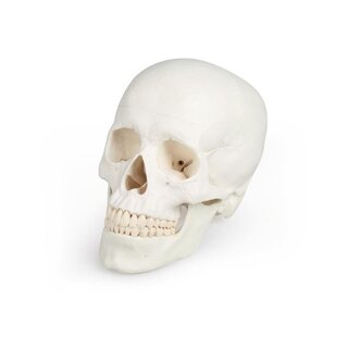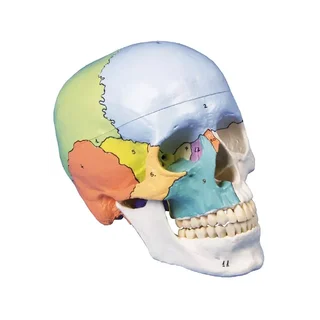


Skull with masticatory muscles, two parts
The masticatory muscles (masseter, temporal, medial and lateral masticatory muscles) are represented as elastic bands. This model is designed to demonstrate the function of the masticatory muscles in relation to jaw closure, initial jaw opening and …
 Part-pay from 51.77 €/mo
- In stock – delivery 7-10 days
- Cheapest shipping from 11.16 €
- Pay with Swish, card or invoice
- Personal customer service by phone and e-mail
- Swedish online store since 2008
The skull model is developed to demonstrate the movement of the jaw using elastic bands that represent the masticatory muscles. The model is cast in robust plastic and is delivered in a size that corresponds to an adult. The dimensions are 23.2 x 16.7 x 16.7 cm and the weight is approximately 900 grams. The top of the skull is removable so that, among other things, the base of the skull (basis carnii externa) can be studied.
Anatomically, it should be emphasized that this variant of the skull model offers an educational opportunity to study the masticatory muscles and their attachment to the skull. The muscles that can be seen are:
M. masseter
M. temporalis
M. pterygoideus lateralis
M. pterygoideus medialis
The four masticatory muscles are represented on both sides of the skull model with the help of elastic bands and the names of the muscles are even written on the attachments (see pictures on the left). Read more about the mobility below.
In general, a human skull can be divided into two parts and the skull model therefore shows the following:
1) The braincase (neurocranium) which encloses the brain and the organ of hearing and balance.
2) The facial skeleton (viscerocranium) which surrounds the nasal cavity and forms the tooth-bearing framework around the oral cavity. The 32 teeth are also visible.
The braincase consists of eight bones – four unpaired (frontal, ethmoid, sphenoid and occipital bones) and two paired (parietal and temporal bones). All of these bones and their sutures can be identified on the model.
The facial skeleton consists of six paired bones (maxilla, palate, hyoid, nasal and lacrimal bones and the inferior nasal concha), as well as three unpaired bones (mandible, ethmoid and pharynx – some do not include the ethmoid in the facial skeleton). All of these bones and their sutures can also be seen on the skull model. NOTE: The hyoid bone is also included in the facial skeleton but cannot be seen on this model.
The human skull has quite a few holes and channels that contain blood vessels and nerves. Many of the most important holes and channels are visible on this skull model, but not all. The most important anatomical “landmarks” are visible, such as the styloid process, but minor details are not included.
In terms of movement, this skull model is developed to be able to demonstrate the function of the masticatory muscles, thus the following can be demonstrated:
Closing the jaw
Beginning of opening the jaw
Movement of the jaw forward and to the side
Clinically, the skull model can be used to understand many diseases and conditions in the temporomandibular joint and masticatory muscles, such as tension in the jaw and temporomandibular dysfunction (TMD). The model is also good to use for understanding diseases and conditions that can occur in this part of the skeleton.
| Item number | 1020169 |