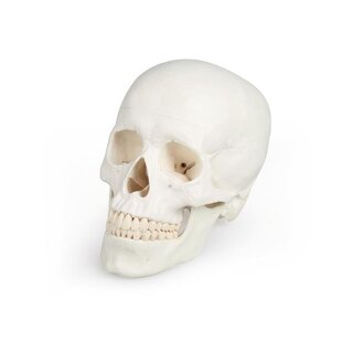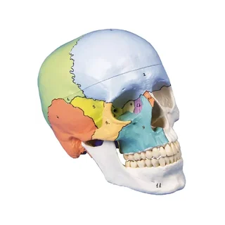


Skull model with blood vessels and nerves in the lower jaw and muscle markings.
This skull model offers a very educational and detailed insight into the blood vessels and nerves of the lower jaw.
 Part-pay from 45.41 €/mo
- In stock – delivery 4-8 days
- Cheapest shipping from 11.20 €
- Pay with Swish, card or invoice
- Personal customer service by phone and e-mail
- Swedish online store since 2008
This skull model offers a very educational and detailed insight into the blood vessels and nerves of the lower jaw. In addition, the following can be seen painted on the skull: the origin and attachment of the muscles, the sutures between the cranial bones, and the arteries and veins on the inside of the cranium.
The model is cast in plastic and is delivered in a size that corresponds to an adult. The dimensions are 20 x 13.5 x 15.5 cm and the weight is approximately 900 grams. The top of the skull is removable so that, among other things, the base of the skull (basis cranii interna) can be studied. The lower jaw bone (os mandibulare) can be easily detached from the rest of the skeleton. Thus, the model can be divided into 3 parts.
The skull model is developed to be able to contain a model of a brain from the same manufacturer. The brain model, which can be divided into five parts, can be seen and purchased HERE.
Both bones, the colored muscle markings, the colored sutures and the lines on the inside of the skull that symbolize blood vessels are numbered on the model. An overview with the names of the different structures is not included with the purchase, but you can download and print it via the link on the left under the product images (click on the PDF symbol). Names are available in Latin, English and several other languages.
Anatomically, it should be emphasized that this skull provides a detailed and educational insight into the blood vessels and nerves of the lower jaw. Anatomical details such as the inferior dental plexus and the a. and v. mentalis can be studied (see images on the left).
In general, the human skull can be divided into 2 parts and the skull model therefore shows the following:
1) The skull (neurocranium) that encloses the brain and the organ of hearing and balance.
2) The facial skeleton (viscerocranium) which surrounds the nasal cavity and forms the tooth-bearing framework around the oral cavity. The 32 teeth are also visible.
The braincase consists of eight bones – four unpaired (frontal, ethmoid, sphenoid and occipital bones) and two paired (parietal and temporal bones). All of these bones and their sutures can be identified on the model.
The facial skeleton consists of six paired bones (maxilla, palate, hyoid, nasal and lacrimal bones and the inferior nasal concha), as well as three unpaired bones (mandible, ethmoid and pluvial bones). All of these bones and their sutures can also be seen on the skull model. NOTE: The hyoid bone is also included in the facial skeleton but is not visible on this model.
A skull has many holes and channels that contain blood vessels and nerves. Many of the most important holes and channels are visible on this model, but not all. The level of detail on the bones is high and many anatomical details are visible, for example the styloid process, but some smaller details are not shown.
The origin and attachment of the skeletal muscles are shown in red and blue on the skull model. These color markings are seen for facial muscles, masticatory muscles, palate muscles, tongue muscles as well as some deep neck muscles and some back muscles.
Sutures are seen in orange and on the inside of the braincase, “meningeal” arteries and veins and venous sinuses are seen with red and blue lines/dashes, respectively.
In terms of movement, it is only relevant to mention the temporomandibular joint. The lower jaw bone (os mandibularis) is held in place by a relatively long metal spring that is attached to the lower and upper jaw bones via screws. Large rubber pieces are mounted on the mandibular fossa (fossa mandibularis) of the jaw joint, which hold the joint head (caput mandible) in place as if in a “rubber bowl” – see pictures on the left.
The model’s jaw joints are not particularly mobile. The lower jaw can be moved up and down and to the sides, but the sliding joint mechanism that the jaw joint normally has cannot be demonstrated.
Clinically, the skull model is perfect for understanding the diseases and conditions that can occur in this part of the skeleton.
| Item number | 1000054 |