

Flexible model of the spine with nerves, hip joint and tripod
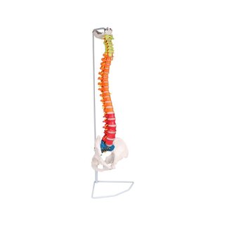
Flexible model of the spine with nerves, hip joint and tripod.
 Part-pay from 24 €/mo
- In stock – delivery 4-8 days
- Cheapest shipping from 26.78 €
- Pay with Swish, card or invoice
- Personal customer service by phone and e-mail
- Swedish online store since 2008
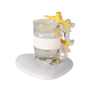
This model of the spine is very flexible and shows the vertebral artery, spinal nerves, neck bone, pelvis, hip joint and a herniated disc.
The model measures 78 cm in height. All vertebrae are held together by a metal tube, which is usually only found in more advanced spine models. In addition, an elastic system has been mounted in the upper cervical vertebrae. The model is supplied with a white metal stand to hang on.
Anatomically
Anatomically, the model shows the entire vertebral column, which consists of a neck, a thoracic section, a lumbar section, a pelvic section and the coccyx. The neck bone is also included. Therefore, the model shows the two most important types of joints in the spine, which are the symphysis (discs/ribs) between the vertebral bodies and the facet joints, which are sliding joints between the pivots of the vertebrae. The lower cervical joint and the sacrococcygeal joint are also visible. Since the neck bone is included in this model, the upper neck joint is also visible.
In addition, the model shows the pelvis in its entirety as a ring structure as well as both the femoral heads with the femoral neck and part of the femur. Therefore, the model also shows the SI joint between the sacrum and the pubic bone, the symphysis between the pubic bones and the hip joint.
The model also shows the vertebral artery, the 31 pairs of spinal nerves and a herniated disc in the lower back. Overall, the bone quality is very good and therefore, for example, knots and depressions in the bone tissue are clearly visible.
Movement
In terms of movement, the model is very flexible. The mobility of the human spine is greatest in the cervical and lumbar spine (which are lordotic), while the thoracic region (which is kyphotic) is less mobile. This model can be used to demonstrate the most important movements of the spine, which are flexion-extension, lateral flexion and rotation in the neck and lumbar spine.
The above-mentioned metal tubes ensure both that you can detach the disc from the vertebrae without it falling apart, and that stability is felt when moving the spine.
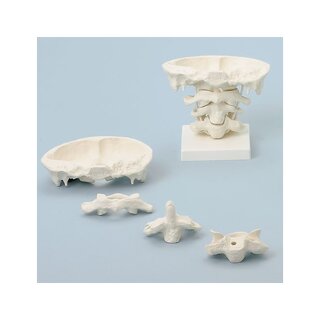
The occipital bone (os occipitale), the upper cervical vertebra (atlas) and the second upper cervical vertebra (shaft) are joined together with an elastic cord, which gives a special elastic effect, where the vertebrae both follow each other during movement and come together again in a natural way. You can thus demonstrate the most important movements of the neck joints, which are the nodding and rocking of the head, which occurs in the upper cervical joint, and shaking of the head, which occurs in the lower cervical joint.
In the hip joints, the femoral head is held firmly in the hole via elastic. Therefore, various movements of the hip joint, such as abduction and rotation, can be demonstrated. The femoral head can also be completely removed.
Clinically
Clinically, this model is ideal for understanding disc herniation, root impingement, and spondylolisthesis. It can also be used to understand diseases such as back pain, scoliosis, spondylosis, spinal stenosis, and osteoarthritis.
The model can of course also be used to understand fractures, vertebral artery dissection, and many different disorders related to the hip joint and pelvis.
| Item number | A123S |
| EAN/GTIN | 5744004481407 |