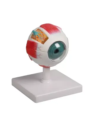
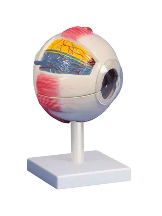
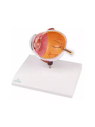
Eye in orbit with eyelid
Anatomical model Eye in orbit with eyelids, 13 parts Approx. 2x enlarged This detailed and approximately twice enlarged model shows the eye placed in the eye socket (orbita) together with surrounding structures and eyelids.

- In stock – delivery 4-8 days
- Cheapest shipping from 8.85 €
- Pay with Swish, card or invoice
- Personal customer service by phone and e-mail
- Swedish online store since 2008
Anatomical model Eye in orbit with eyelids, 13 parts
Approx. 2x enlarged
This detailed and approximately twice enlarged model shows the eye placed in the eye socket (orbita) together with surrounding structures and eyelids. The model consists of 13 separate parts and is designed to provide a complete and educational overview of both the internal components of the eye and its relationship to the surrounding tissue.
It is ideal for teaching anatomy, ophthalmology, optometry and medical education as well as for visual patient information in clinical practice.
The model shows, among other things:
- Eyeball with removable components: cornea, iris, lens, vitreous humor, etc.
- Eyelid with internal structure
- Eye muscles in correct position
- Optical nerve and blood vessels
- Bone structures in the eye socket (orbita)
- Fatty tissue and supporting soft tissues
All parts are hand-painted in lifelike colors and can be disassembled to enable detailed study of each individual component.
Features:
Magnified approximately 2 times for increased clarity
13 parts detachable for interactive teaching
Anatomically correct and realistically colored
Shows both internal eye structure and surrounding tissue
Mounted on base for stability
Perfect for:
University and school teaching
Education of ophthalmologists and opticians
Patient consultation and visual explanation in healthcare settings
| Item number | F160 |
| EAN/GTIN | 4250395323055 |