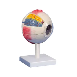
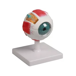
Half of the eye, enlarged
Cross-section model of the eyeball - detailed overview of the internal anatomy of the eye This anatomical model shows a carefully reproduced cross-section through the eyeball, providing a clear insight into the internal structures of the eye.
 Part-pay from 24.47 €/mo
- In stock – delivery 4-8 days
- Cheapest shipping from 8.87 €
- Pay with Swish, card or invoice
- Personal customer service by phone and e-mail
- Swedish online store since 2008
Cross-section model of the eyeball - detailed overview of the internal anatomy of the eye
This anatomical model shows a carefully reproduced cross-section through the eyeball, providing a clear insight into the internal structures of the eye. The model is designed to illustrate both the optical components and the neurovascular and muscular parts of the eye.
The following anatomical structures are depicted:
- Sclera
- Cornea
- Iris and lens
- Ora serrata
- Macula
- Retina
- Choroidea
- Optical nerve (nervus opticus)
- Central retinal artery and vein
- Retinal blood vessels
- Superior and inferior rectus muscles (eye muscles)
The model is excellent for teaching anatomy and eye care, as well as as an educational tool for opticians and ophthalmologists when providing patient information. It makes it easy to explain the function of the eye and common vision-related conditions in a visual and easy-to-understand way.
Features:
Realistic and detailed cross-sectional model
Shows both structures and vascular supply
Ideal for education, demonstration and advice
Suitable for schools, universities, opticians and healthcare practices
A compact and clear model that provides a complete overview of the anatomy of the eye, perfect for both professional and educational use.
| Item number | F50 |
| EAN/GTIN | 4250395320009 |