

Foot with muscles, blood vessels, nerves, ligaments in six parts
Foot with muscles, blood vessels, nerves, ligaments in six parts This anatomically detailed model of the foot and lower leg can be separated into six parts for a detailed examination of the foot and ankle.
 Part-pay from 40.60 €/mo
- In stock – delivery 2-3 weeks
- Cheapest shipping from 11.15 €
- Pay with Swish, card or invoice
- Personal customer service by phone and e-mail
- Swedish online store since 2008
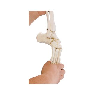
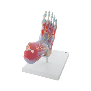
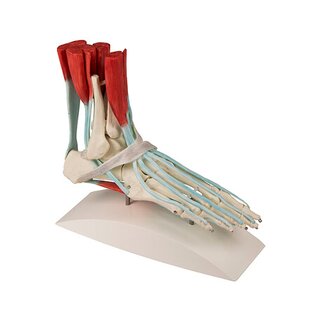
This anatomically detailed model of the foot and lower leg can be divided into six individual parts, which allows for a detailed examination of the foot and ankle. The foot skeleton demonstrates not only the bones but also the muscles, tendons, ligaments, nerves, arteries and veins of the foot.
The extensor muscles of the legs can be seen on the foot model. The tendons can be followed in their passage under the transverse and vertical ligaments, all the way to the console. In addition, all the sensor feet on the foot can be seen. On the dorsal side of the foot, the gastrocnemius muscle can be removed. The foot of the foot is represented in three layers: the first layer shows the flexor digitorum brevis. This muscle can be removed to visualize the quadratus plantar, the tendon of the flexor digitorum longus and the flexor hallucis. The second layer is in turn also removable to show even deeper anatomical details of the foot. The model is of very high quality and the buyer gets a lot for their money.Anatomically, the model contains various structures such as bones, ligaments, muscles, tendons, arteries and nerves in both the ankle segment, on the ankle, in the sole of the foot and in the toes.
The foot model really contains many anatomical structures - for example the following:
The Achilles tendon and a cross-section of the muscles of the lower leg (above the ankle region).
Furthermore, you can follow the attachments of these muscles in the foot in an educational way
Tarsal tunnel under the flexor retinal axis on the inside of the ankle containing tendons, vessels and nerves
Muscles, tendons, nerves and blood vessels on the back of the foot
The superficial plantar aponeurosis (fascia plantaris) as well as the muscles of the sole of the foot which are divided into respectively. the great toe lodge, the central lodge, the small toe lodge and the spatia interossea
Bones, ligaments, tendons, vessels and nerves of the toes (distributed)
In terms of movement, the model is not flexible. The model cannot be used to show movements of the ankle.
Clinically, the model is ideal for understanding disorders such as Achilles tendon rupture, heel spurs / plantar fasciitis and tarsal tunnel syndrome.
This complete model is also ideal for understanding toe fractures, luxations (joint defects) in the toes and foot abnormalities such as hallux valgus, hallux rigidus, hammertoe, flat feet, clubfoot and metatarsus varus.
The model can also be used to understand disorders such as osteoarthritis of the foot (osteoarthritis) and rheumatoid arthritis (rheumatoid arthritis).
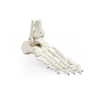
NOTE: If you are looking for a complete foot model that can be separated into even more parts, we recommend our other complete foot model. On this model, the anatomical structures are numbered and named in the accompanying overview.
| Item number | 1000360 |