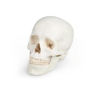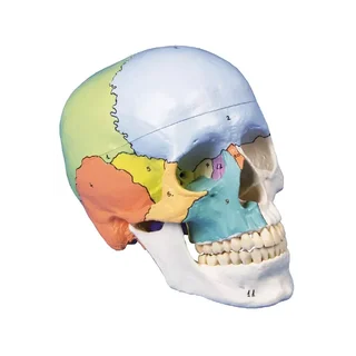


Color model of fetal skull at week 38
This model of a fetal skull comes with educationally colored skull bones that also highlight the fontanelles. The fetal skull is cast in colored plastic and comes in a size that corresponds to the size of the fetus in the 38th week of pregnancy.
 Part-pay from 18.36 €/mo
- In stock – delivery 4-8 days
- Cheapest shipping from 11.20 €
- Pay with Swish, card or invoice
- Personal customer service by phone and e-mail
- Swedish online store since 2008
This model of a fetal skull comes with educationally colored skull bones that also highlight the fontanelles.
The fetal skull is cast in colored plastic and comes in a size that corresponds to the size of the fetus in the 38th week of pregnancy. The weight is 0.2 kg. Both the colored skull bones and the fontanelles are numbered. A list with the names of these anatomical structures is included in the purchase.
Anatomically, it should be emphasized that the fetal skull provides the opportunity to see the fontanelles. Although a child has 6 of these areas (holes) at birth, it is often only spoken of as an anterior and a posterior fontanelle.
All 6 fontanelles are visible on the fetal skull;
The large anterior fontanelle (seen in color on the model)
The posterior fontanelle
Right anterior lateral fontanelle
Left anterior lateral fontanelle
Right posterior lateral fontanelle
Left posterior lateral fontanelle
Generally speaking, a human skull can be divided into two parts and the fetal skull model therefore shows the following:
1) The braincase (neurocranium) which encloses the brain and the organ of hearing and balance.
2) The facial skeleton (viscerocranium) which surrounds the nasal cavity and forms the tooth-bearing framework around the oral cavity. The 32 teeth are also seen.
The different cranial bones are of different colors, but paired bones are of the same color.
In terms of movement, it is only relevant to mention the temporomandibular joint, which is not movable on this model.
Clinically, the model is developed to understand fontanelles, which often concern new parents. The model can also be used to understand conditions such as craniosynostosis and the formation of independent suture bones (ossa suturalia/Wormiana) as well as the development and function of sutures in relation to treatment modalities such as osteopathy and craniosacral therapy.
| Item number | A156 |