Meiosis model
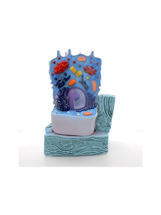
This newly developed model series demonstrates the ten stages of meiosis inside a typical mammalian cell at a magnification of approximately ten thousand times. 1. Interphase (stage of G1 phase) 2. Prophase I (leptotene) 3. Prophase I (zygotene

- In stock – delivery 2-3 weeks
- Cheapest shipping from 8.85 €
- Pay with Swish, card or invoice
- Personal customer service by phone and e-mail
- Swedish online store since 2008
This newly developed model series demonstrates the ten stages of meiosis inside a typical mammalian cell at a magnification of approximately ten thousand times. 1. Interphase (stage of G1 phase) 2. Prophase I (leptotene) 3. Prophase I (zygotene and pachytene) 4. Prophase I (diplotene) 5. Prophase I (diakinesis) 6. Metaphase I 7. Anaphase I 8. Telophase I, cytokinesis I, interkinesis, prophase II and metaphase phase II 9. Anaphase II 10. Telophase II and cytokinesis II. The three-dimensional relief model is colored according to standard microscopic methods, which makes meiotic cell division easy to understand. The cell's organelles are displayed openly in the lower part of the model. The model is equipped with magnets so that it can be easily placed on a magnetic board in the classroom. The model series is also included in a storage system (40 x 60 cm) that can be hung on a wall. Comes with a detailed description and stencils that can be handed out in lessons. Tip: As a great addition and eye-catcher in the classroom, we recommend the matching mitosis poster (V2051U).
| Item number | 1013869 |